

Physiochemical properties such as solubility and dissolution rate play a crucial role when it comes to the therapeutic efficacy of oral drug formulations. Most drug candidates exhibit modest water solubility and low intestinal permeability,1 resulting in poor absorption and an insufficient concentration of the drug in the body.
To bring such drugs to the market, their physiochemical properties must first be improved. One approach involves modification of the drug’s molecule; however, that may lead to undesirable therapeutic effects or adverse side effects.2
Crystal engineering is an approach that allows scientists to tune the physical properties of pharmaceutical materials in the solid form through deliberate control of intermolecular interactions between the molecules of the active pharmaceutical ingredient (API) while still retaining their original chemical functionality. This manipulation of APIs is often accomplished through the design and formation of polymorphs, salts, hydrates, solvates, and co-crystals.
For example, the drugs valsartan and sacubitril are used to regulate blood pressure. Co-crystallization of both drugs in the presence of sodium cations results in a new crystalline phase (patented as Entresto® by Novartis) with 50% higher bioavailability of valsartan compared to the valsartan administered alone.3
Many crystal engineering methods are scalable and simple to implement: for instance, solid-state grinding, solution-reaction crystallization, solvent evaporation, slurry conversion, and hot-melt extrusion.4 Reaction conditions can be controlled and optimized to attain the desired purity of the final material. Given the number of experimental variables at play, discovery of new drug formulations and optimization of experimental conditions for their synthesis are usually done in a high-throughput fashion.
High-throughput powder x-ray diffraction (HT-PXRD) allows for characterization of hundreds of samples with a single well plate in a matter of hours. A refined micro-x-ray beam moves across the wells continuously, probing various regions of the sample (see figure to the right).
Crystalline powder samples diffract x-rays in accordance with Bragg’s law, forming diffraction peaks that are recorded by a detector installed on a goniometer.
Diffraction peaks (and the resulting diffraction patterns they form) contain information about the crystal structures present in the sample. By comparing measured diffraction patterns with database entries, one can identify the presence of unreacted initial reagents, formed by-products, and new phases that have yet to be reported in the database. Given the distinctiveness of diffraction patterns, all observed peaks from the collected data should match with the peaks from the database entry to confirm the presence of a particular phase. This is why this analytical technique is called fingerprinting.
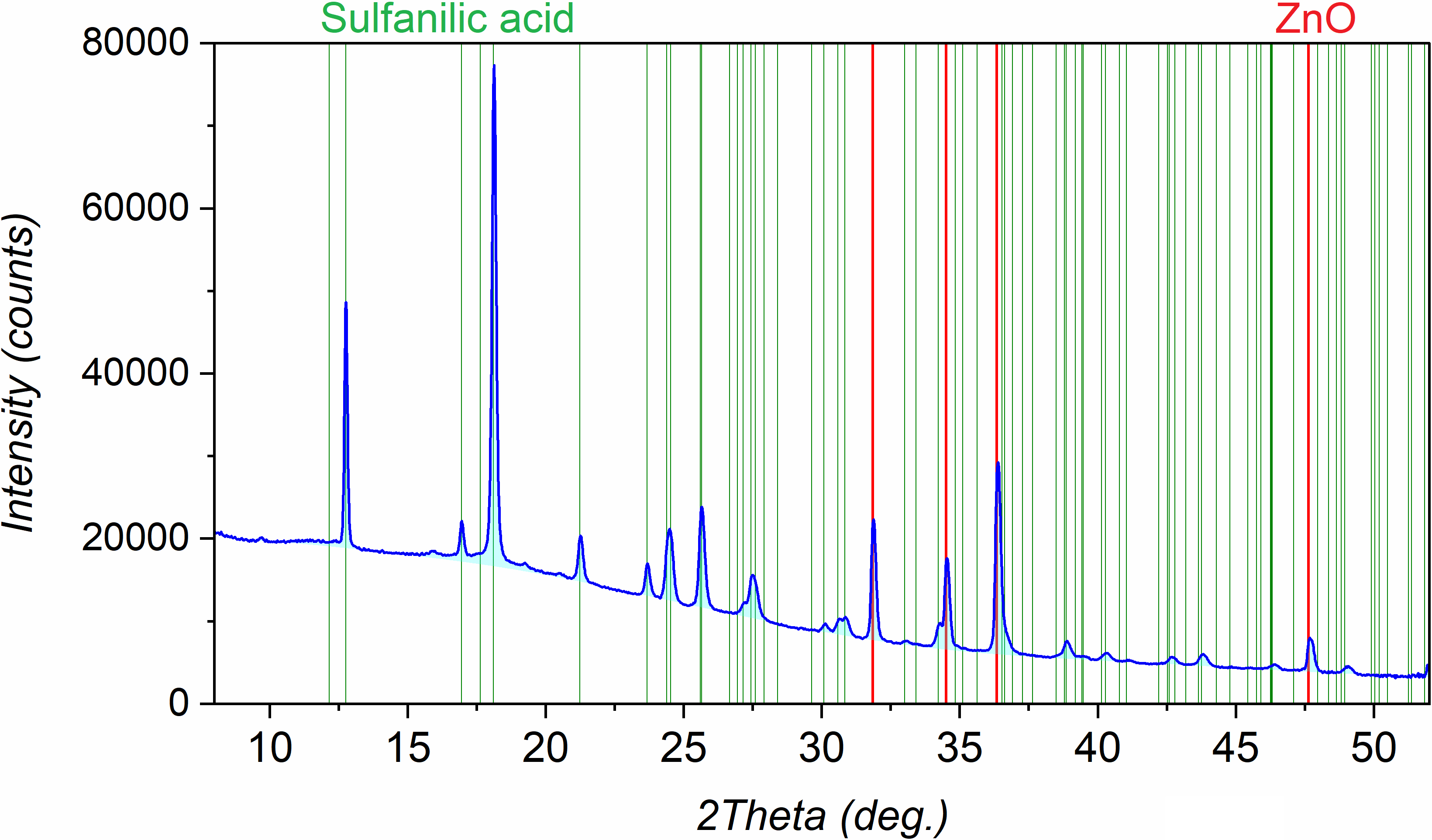
Consider the PXRD pattern to the right. A thorough search across entries in the COD and CCDC databases revealed the presence of two phases: sulfanilic acid and zinc oxide.
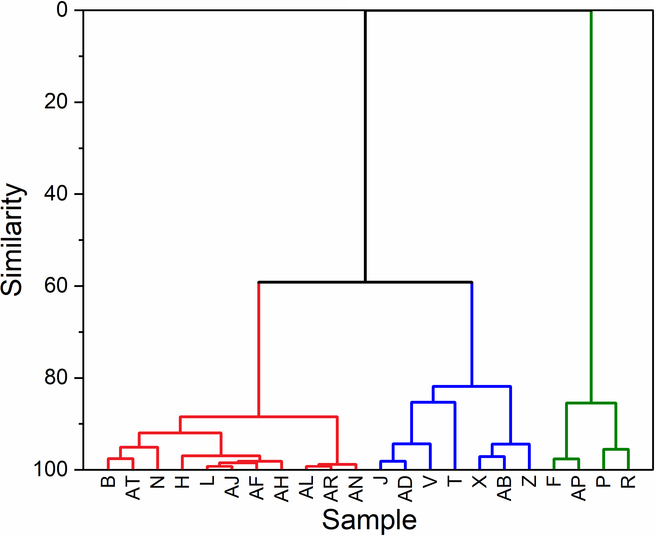
To expedite analysis of the large number of diffraction patterns collected during high-throughput screening, cluster analysis can be employed. Comparing all datasets point by point, the algorithm can reveal similar patterns and group them together. Identifying common traits in the group allows one to extrapolate the results to all samples within the same group.
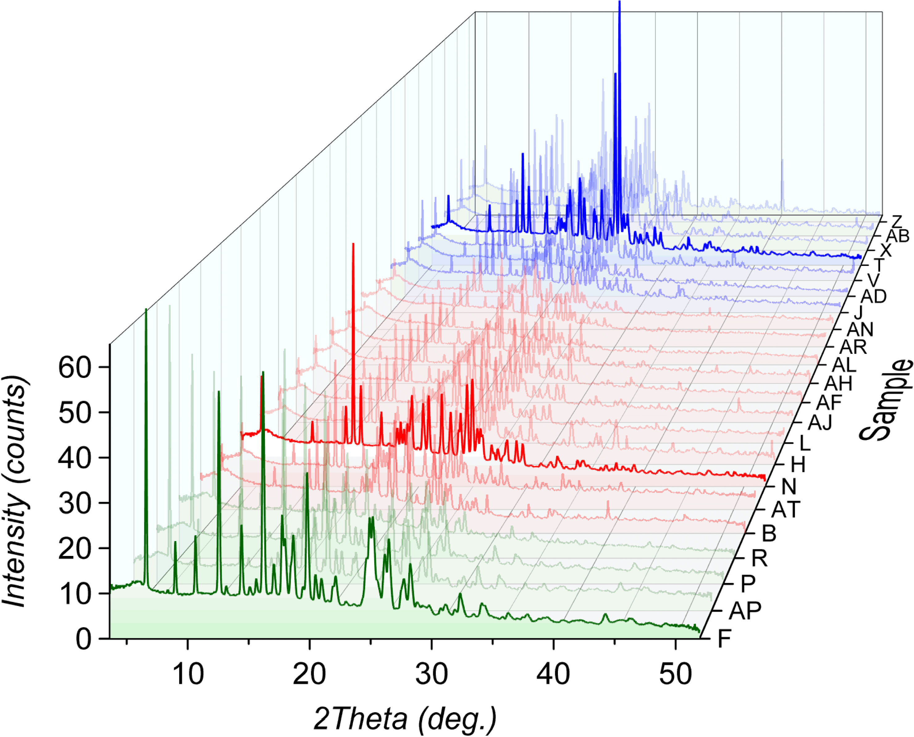
The following dendrogram was plotted for 22 samples obtained after mechanochemical optimization of a co-crystal formation. Every sample was prepared by using the same API and the same co-former; however, the solvent used for the synthesis was different for each sample. Cluster analysis shows three distinctive groups of patterns (red, blue, and green). While the red and blue groups belong to the same clade (or branch), the green group stands out. Further phase analysis confirmed the presence of a new co-crystal phase in the green group, while the samples identified as the red and blue groups were a mixture of initial reactants.
When considering an HT-PXRD system for a pharmaceutical lab, scientists should be aware of some key elements that will help ensure optimal results.
• Transmission geometry: While working with weakly absorbing samples (organic small molecules and proteins), transmission geometry maximizes diffraction signal by letting the x-ray beam travel through the bulk of the sample, dramatically increasing the sample-illumination volume.
• Copper anode: The longer wavelength of copper (compared to other common anode materials like Mo or Ag) is less penetrative and therefore better interacts with light scatterers, which increases the intensity of the diffraction signal. Additionally, longer wavelength results in better peak separation, which facilitates the analysis of molecular samples in which the number of peaks is especially high (due to low symmetry).
• Microfocus and x-ray optics: A microfocus x-ray source can increase the brightness of the incident beam by more than 10 times compared to traditional sealed x-ray tubes. Microfocus x-ray tubes enable the use of Montel optics, which further refine the beam, eliminating the contribution of the Kβ x-ray component and focusing the beam at the surface of the detector to minimize peak broadening.
• Hybrid photon-counting detectors: Detectors that utilize hybrid photon-counting technology provide low background and high efficiency of signal capture. Large area detectors can be considered to simultaneously collect intensities from a wide range of 2theta angles.
• Well plate technology: Large well plates enable the characterization of many samples without user intervention. Proto’s AXRD LPD-HT comes with a well plate holder that is compatible with most commercially available well plates, allowing for the collection of hundreds of samples with just one well plate.
• Extended functionality: Additional in-situ stages, such as variable temperature/pressure units or humidity chambers, enable expanded testing of the stability of new drug formulations.


